A three-part installation consisting of a striking 16th-century portrait of the notorious Tudor monarch Henry VIII, alongside two monitors with ultra-high resolution scanned reproductions of the original painting.
Detail of ultra-high-resolution scan by LuxLab, UNSW Sydney, Australia

When the Portrait of Henry VIII was examined in 2014, it was also imaged using ground-breaking ultra-high-resolution scanning technologies at 1200dpi at LuxLab, UNSW Sydney. The LuxLab system incorporates a trichromatic CCD camera, macro lens and scanner controller modules developed by UNSW’s Advanced Imaging Laboratory. The resulting image can be digitally magnified many times the size of the original object, as evidenced on the touchscreen display in Henry VIII Trifold. At higher levels of magnification, cracks, areas of loss, brush marks and traces of paint become apparent that are normally invisible for humans, such as the tiny stroke of blue paint in Henry VIII’s right eye.
The holistic comparison of the XRF maps and LuxLab scans thus reveals new insights. For example, examination of the sitter’s right eye in both the scan and XRF map pinpoints the presence of a tiny stroke of copper- based blue, approximately one millimetre in length, therefore presumed to contain the pigment azurite. No other use of blue pigments has been identified anywhere else on the painting. This has provided a further link to the National Portrait Gallery Henry VIII, in which azurite has also been similarly identified in the eye.
Portrait of Henry VIII Attributed to an anonymous Anglo- Flemish workshop, 1535–1540, oil on oak panel, 54.5 x 38 cm. Collection Art Gallery of New South Wales, Australia, purchased 1961. Pre-restoration image, courtesy Art Gallery of New South Wales.

One of the most prominent features of this portrait of Henry VIII is its tendency to recall the more famous portrait by Hans Holbein the Younger. This painting is in fact one of dozens of similar representations created during King Henry’s reign. In a precursor to mass industrial reproduction, portrait artists generated copies by tracing face patterns, which were thought to have been shared among artists and workshops, to mark the position of the sitter and their features. The following text is sourced from the 2015 scholarly article ‘Mapping Henry’ (Dredge et al. 2015), produced by the collaborators that undertook the high resolution scanning an analysis of the work in 2014.

In 2014, prior to planned conservation treatment, the Portrait of Henry VIII was examined for the first time using high-definition X-ray fluorescence (XRF). A primary objective of this exercise was to compare Sydney’s Henry VIII with four similar early Tudor portraits of the King held in British collections, in partnership with a research project led by London’s National Portrait Gallery. The informative and detailed elemental maps, alongside ultra-high-definition scans of the painting undertaken before and after varnish and over-paint removal, have assisted in comparison of the finely painted details with the London paintings. All five paintings share compositional features, although none of them is an exact copy of the other, with differences in costume details, the position of the fingers, and the measurements of each panel.
The Sydney Henry VIII painting is an excellent case study for XRF mapping as it utilises a small number of pigments: copper-based green, mercury-based vermillion, iron- and manganese-based oxide browns, calcium-based ivory black, gold, lead white and chalk. Thus, the distribution of almost all the paint layers can be examined by their elemental fingerprint.
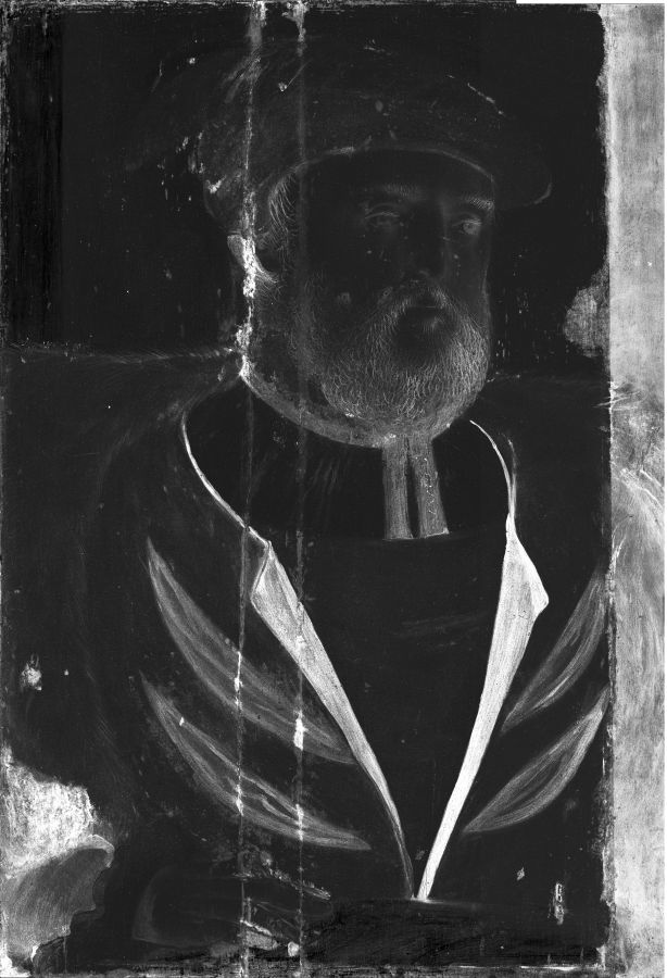
Several new discoveries about the making of the painting are disclosed by the XRF map of the distribution of lead-based pigments. A comparison of techniques of applying the lead white ground layer in a diagonal pattern may be an important clue to a shared workshop practice if the ground application on the London painting was to be similarly imaged. The brush strokes used to apply the priming radiate diagonally across the panel from a starting point at the upper right and describe a brush width of approximately 2.5 cm. Similar diagonal brush strokes in the lead white priming layer are described as texturally visible ridges on parts of the Henry VIII painting at the National Portrait Gallery, London. The detail of a hidden slashed sleeve of the right proper arm is also revealed in the XRF lead map of the Sydney painting, despite being obscured by a broad area of brown restoration paint.
The narrow vertical passage of green paint extending down the right edge of the panel from top to Henry’s proper left shoulder was shown by XRF mapping to be principally a chromium-based green pigment. As the metal chromium was not known until its discovery in 1797 and did not appear in pigments on artist’s palette until the nineteenth century, this cannot be original.
The information seen in the XRF gold map of the painting additionally provides an extraordinary documentation of gold leaf technology of the period. Crease and fold lines created during the gilding process are visible as sharp fine lines, but visible too are the more diffuse variations in the gold thickness, called lamellae, created during the beating of the gold into thin leaves.